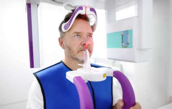

Advanced dental imaging has become an important part of modern dentistry, particularly when a detailed view of the teeth and jaw is needed. A Dental CBCT Scan in Dubai uses cone beam computed tomography to produce three-dimensional images of dental and facial structures. These images can provide information that may not be visible through a standard dental X-ray.
While CBCT technology is highly useful, safety remains an important consideration. The scan uses ionizing radiation, so dental professionals generally recommend it when the additional information is clinically valuable. The goal is to obtain the necessary diagnostic information while keeping radiation exposure as low as reasonably achievable.
What Makes CBCT Different?
Traditional dental X-rays typically create two-dimensional images, while CBCT produces a three-dimensional representation of the scanned area. This allows dental professionals to examine teeth, roots, jawbone, and surrounding anatomical structures from different angles.
The additional detail can be particularly useful for complicated cases. For example, a dentist may require a more comprehensive view before placing an implant, removing an impacted wisdom tooth, or investigating unusual tooth-root anatomy.
CBCT does not replace conventional dental X-rays in every situation. Instead, it serves as an advanced diagnostic option when a three-dimensional view can contribute meaningful information to diagnosis or treatment planning.
Radiation Exposure and Patient Safety
Radiation exposure is one of the main factors considered before any imaging examination. CBCT generally exposes a patient to more radiation than a routine intraoral dental X-ray, although the actual dose can vary considerably depending on the machine, field of view, and imaging settings.
A dental professional should therefore determine whether CBCT is appropriate for the specific situation. When a scan is necessary, modern equipment and appropriate imaging protocols can help limit unnecessary exposure.
The smallest suitable scanning area may be selected when clinically appropriate. This can allow the dental team to focus the examination on the region that requires assessment rather than imaging a larger area without a specific reason.
How a Dental Clinic Maintains Imaging Accuracy
A Dental Clinic using CBCT technology can incorporate three-dimensional imaging into a broader diagnostic process. The scan is not considered a standalone diagnosis. Instead, dental professionals interpret the images alongside the patient's medical and dental history, symptoms, clinical examination, and other diagnostic findings.
Image quality can depend on several factors, including patient movement, positioning, equipment settings, and the area being examined. Following the technician's instructions during the scan can help produce clearer images.
Accurate imaging is especially important when detailed anatomical information is required for treatment planning. A clear CBCT image can help the dentist evaluate the relationship between teeth, roots, nerves, bone, and other structures.
CBCT for Dental Implant Planning
Dental implant treatment is one of the areas where three-dimensional imaging can provide significant value. Before implant placement, the dental professional may need to assess the amount and shape of available bone and identify important structures near the proposed implant site.
A CBCT scan can provide cross-sectional and three-dimensional views of the jaw. This may help the dentist determine whether the existing bone appears suitable for the planned procedure and identify anatomical considerations that could influence treatment.
The information may also assist with selecting implant positioning and planning procedures such as bone grafting when clinically required.
Accuracy in Root Canal Treatment
Complex root canal anatomy can sometimes be difficult to understand using conventional two-dimensional X-rays. Teeth may have multiple canals, unusual root shapes, or anatomical variations that are not easily visible in standard images.
In selected cases, CBCT imaging can provide additional information about the tooth and surrounding structures. This may help a dental professional investigate persistent symptoms, assess complicated root anatomy, or evaluate certain endodontic problems.
However, CBCT should not automatically be used for every root canal patient. The decision depends on the circumstances and whether the additional information is expected to influence diagnosis or treatment.
Assessing Impacted Teeth
Impacted wisdom teeth and other unerupted teeth can have unusual positions within the jaw. Their proximity to nerves, neighboring teeth, and other structures may be important when planning treatment.
Three-dimensional imaging can show the position of an impacted tooth from multiple perspectives. This can give the dental team a better understanding of the surrounding anatomy before deciding on an appropriate treatment approach.
Such information can be particularly valuable when a tooth appears to have a complicated relationship with nearby structures on conventional X-rays.
Accuracy Depends on Professional Interpretation
Even high-quality imaging requires proper interpretation. A CBCT scan produces a large amount of anatomical information, but the significance of that information must be evaluated by a qualified dental professional.
The images may reveal structures or findings that require further clinical assessment. Therefore, patients should not attempt to interpret a CBCT image on their own based solely on what appears on the screen.
A dentist considers the complete clinical picture before recommending treatment. This approach helps ensure that imaging findings are interpreted in the appropriate context.
When Is a CBCT Scan Recommended?
A CBCT scan may be considered for complex dental implants, impacted teeth, selected root canal cases, oral surgery, orthodontic assessment, jaw evaluation, and other situations where three-dimensional information is useful.
It is not normally required for routine dental checkups or every common dental problem. The appropriate imaging method depends on the patient's condition and the diagnostic question the dentist needs to answer.
Final Thoughts
Safety and accuracy are both important when advanced dental imaging is used. When clinically justified, Dental CBCT Scan technology can provide detailed three-dimensional information that supports diagnosis and treatment planning. Choosing the appropriate scan, using suitable imaging protocols, and having the results professionally interpreted can help make the technology a valuable part of modern dental care.